

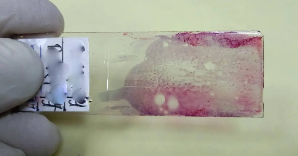
Most pathology reports after pancreatic cancer surgery answer one question: how much tumor is left? A new study from Mayo Clinic suggests that’s only half the story. The shape and arrangement of that remaining tissue may matter just as much, and AI can measure it.
What the researchers found
Published in Clinical Cancer Research, the study analyzed standard H&E pathology slides from 203 patients with pancreatic ductal adenocarcinoma who had received chemotherapy before surgery but showed only a limited response. Using an AI-enabled digital pathology platform combined with methods borrowed from landscape ecology, the team measured how cancer and surrounding stromal tissue were spatially organized. Specifically, they looked at fragmentation, patch boundaries, and how intermixed the two tissue types were.
Patients with more fragmented, intermixed patterns relapsed sooner. Standard measures, including the total amount of residual cancer, did not reliably separate high- and low-risk patients. But the spatial models did. In one model, high-risk patients had a 71% greater adjusted risk of recurrence. In another, their risk was more than double.
Why arrangement beats amount
“Current pathology assessments largely tell us how much tumor is left after treatment,” says Ryan Carr, M.D., Ph.D., Mayo Clinic oncologist and senior author. “We wanted to know whether the geography of that remaining cancer could reveal additional biology about recurrence risk.”
The study also found that high-risk spatial patterns contained fewer immune cells inside the tumor itself. Instead, immune cells tended to cluster around it rather than infiltrate it. That points to the tumor microenvironment as a key driver of treatment resistance, a finding that fits broader oncology research trends.
Clinical relevance and what comes next
Because the analysis runs on slides already generated during routine care, it wouldn’t require additional tissue testing. Still, the researchers are clear: these results need prospective validation before the approach can inform real clinical decisions.
The long-term aim is to use spatial risk profiling to guide surveillance schedules, adjuvant therapy choices, and clinical trial design for patients who remain at highest risk after surgery.